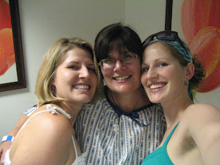
I look adorable in my MRI gown.
Dr. Nino explained that they would try to isolate the same spots seen previously seen on the MRI to biopsy and take a sample from the most promising section. If they couldn't see them then they would not do a biopsy and we would recheck in six months. That seemed like a good scenario to me.

My girls kept me company!
The MRI is done with me laying face down on the MRI table, breasts hanging through a hole. Each breast is squeezed by paddles just like the ones for mammograms except these have gridlines on them and open areas for the biopsy catheters to go through. One MRI is done to see if they can find the spots they saw previously ( they could see them both) and where the spots are in relation to the grid. This took two tries and then they brought me out of the MRI tunnel and cleaned the skin and numbed the area. After a few minutes, Dr. Nino placed the catheters in each breast using the grids to find the correct area. A quick trip back into the MRI machine was done to make sure the catheters were in the right spot and then the tissue samples were taken and titanium clips placed to make it possible for the surgeon or a future radiologist to find the exact area at a later time.
After the procedure a mammogram is done, ice applied. There is a lot of bruising and more discomfort after this biopsy because the MRI identifies areas that are needing more blood supply so there is more blood in the areas they sampled than the last biopsy.
Results sometime Monday!

